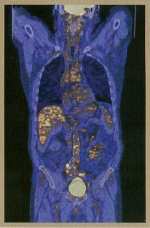
Positron Emission Tomography(P.E.T.)

A positron emission tomography (PET) scan is a unique noninvasive imaging technique that can produce three-dimensional images of the living heart, brain or other organs at work.
PET scans are often used in the diagnosis and management of cancers, certain brain disorders and heart disease. Cardiac PET scanning is generally similar to other types of non-invasive stress tests to help determine the presence and extent of CAD. It has two major advantages over the more common nuclear stress tests . First, the images are less likely to be distorted by parts of the patient's body (large breasts, obesity etc.), so abnormal results are more reliable. Second, it is an excellent tool for determining whether portions of the heart muscle are still viable (living and functioning). The scan can also measure how well those viable portions are functioning after a heart attack or other event in which there is a lack of oxygen-rich blood to the heart muscle.
PET scanning is now as readily available as more conventional nuclear imaging because of the construction of cyclotron devices, which produces necessary isotopes.